


Quibim has launched QP-Breast, an AI-driven decision-support device which automatically identifies lesions doubtful for breast cancer on MRI, making it the first such device to receive both CE and UKCA design in Europe and the UK. The tool is CE marked under EU MDR 2017/745 as Class IIb and UKCA marked as Class IIa.
QP-Breast assists radiologists by highlighting doubtful regions and generating structured reports detailing lesion size, volume, location, and likelihood of malignancy, integrating directly into presenting hospital technology. The device is intended for both screening and diagnostic breast MRI, aimed at hospitals, screening programmes, and specialist imaging centres.
The launch responds to increasing requests for radiology services, as breast cancer remains the most generally diagnosed cancer in women worldwide, with 2.3 million new cases yearly, while up to four in five biopsied lesions turn out not to be cancer, driving unnecessary measures and expenses.
QP-Breast follows Quibim's flagship QP-Prostate, which holds CE, UKCA, and FDA 510(k) clearance. More than 470 sites globally have applied Quibim's imaging products.
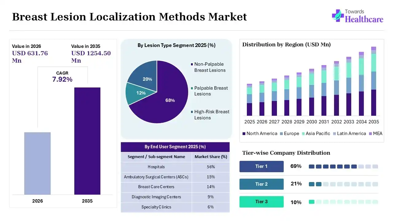
According to Towards Healthcare, the breast lesion localization methods market is projected to experience significant growth, with estimates suggesting the market size will increase from USD 631.76 million in 2026 to approximately USD 1254.5 million by 2035. Estimated to grow at a CAGR of 7.92% starting from 2026 to 2035 Breast lesion localization method involves implanting a small tag into the breast lesion before surgery. This is completed by a radiologist under ultrasound or X-ray guidance. Every tag contains a specific identifier and emits a radio-frequency signal noticeable by a handheld reader. This process provides real-time identification and enables placement days in advance, improving flexibility for both patients and surgical leaders. Breast lesion localization processes have progressed to more patient-friendly technology, each with its unique advantages. Future research on patient-centric results, cosmetic outcomes, breast biopsy markers, and incorporation of augmented reality with breast lesion localization is required. It improves surgical precision, patient safety, and comfort, while also optimizing hospital resources.
Quibim, a worldwide leader in whole-body automated healthcare imaging analysis, follows an AI-driven approach to support identifying pathologies in each body part and imaging modality. This organization believes that evidence-based research and technological advancement, integrated, support saving lives. Quibim is an organization designing and creating pioneering tools that unlock imaging data to increase drug programmes and enhance patient results. The organization uses imaging modalities such as MRI, CT, and PET to support detecting pathologies and predicting outcomes in oncology/immunotherapy, rheumatology, and neurology using AI-driven strategies. The main aim is to transform imaging into actionable predictions. Real-world evidence (RWE) and biopharma collaborate to leverage Quibim's expertise in big imaging infrastructures to advance the development of AI models and gain advanced insights from images. Quibim creates advanced quantitative imaging biomarkers to track the effectiveness of treatments and find novel drugs by thoroughly analyzing disease mechanisms. Quibim aims to convert imaging into a catalyst for accurate health by leveraging the strength and strenth of quantified imaging data and AI. To date, Quibim has received over €20 million in public and private subsidies and is a participant in 19 EU projects that have received €30 million in funding.
A recent report by Towards Healthcare highlights that the breast lesion localization methods market is growing as breast irregularity is analyzed; breast cancer localization is often a crucial next step in the treatment technology. This technology plays a significant role in guiding physicians by precisely marking the area of concern before surgery. Breast cancer localization is usually used in technologies like lumpectomy or breast-conserving surgery, where the aim is to remove only the abnormal tissue while preserving as much healthy breast tissue as possible. The localization is continuously performed before surgery, with the timing depending on the particular processes used. Wire-free localization includes placing a small marker right into the abnormal tissue. Unlike wire localization, this process allows the technology to be performed numerous days, or even up to 30 days, beforehand surgery. During the process, the surgeon uses a handheld tool to identify the implanted marker and locate the specific tissue with precision. This strategy provides greater flexibility and enhanced patient comfort.